The Stereo-seq workflow enables researchers to capture and analyze gene expression within tissues while preserving their spatial organization. This approach provides a powerful way to understand how cells interact and function within complex biological structures. The workflow consists of five main stages: sample preparation, staining and imaging, mRNA capture, library construction and sequencing, and computational analysis.
Sample Preparation
The workflow begins with the preparation of fresh frozen tissue sections, which are carefully placed onto a specialized Stereo-seq chip. This chip contains a high-density array of spatially barcoded capture probes that record the precise location of RNA molecules within the tissue. Proper tissue preservation during this stage is critical to maintain both RNA integrity and spatial structure.
Staining and Imaging
After mounting the tissue section, the sample is stained using ssDNA or H&E staining to visualize the tissue morphology. High-resolution imaging is then performed using microscopy. These images serve as an anatomical reference that can later be aligned with spatial gene expression data, allowing researchers to directly link molecular information with tissue structure.
Types and Sizes of Chips
P Chip (Permeabilization Chip): Used to optimize tissue permeabilization conditions prior to the spatial experiment, ensuring efficient RNA release while preserving tissue morphology.
T Chip (Spatial Transcriptomics Chip): Used for fresh-frozen tissue sections, where released mRNA binds to spatially barcoded probes on the chip, enabling high-resolution spatial gene expression mapping.
OMNI Chip (Spatial Transcriptomics Chip): Designed for FFPE (formalin-fixed paraffin-embedded) samples, enabling spatial transcriptomics from archived or clinically preserved tissues.
• 0.5 cm × 0.5 cm T Chip, which is typically used for smaller tissue sections or pilot spatial transcriptomics experiments.
• 1 cm × 1 cm T Chip, which provides a larger capture area and enables profiling of larger tissue sections or multiple tissue regions simultaneously.
• 1 cm × 1 cm Omni Chip v1.1, which supports expanded spatial capture areas and improved experimental flexibility using FFPE samples.
Ask questions for custom chips.
Chip types
Permeabilization and In Situ mRNA Capture
Next, the tissue undergoes permeabilization, which allows messenger RNA (mRNA) molecules inside the cells to diffuse out and bind to the spatially barcoded probes on the chip surface. Each probe carries a unique positional barcode, ensuring that every captured transcript retains information about its original location within the tissue. This step effectively records the spatial distribution of gene expression across the sample.
Library Construction and Sequencing
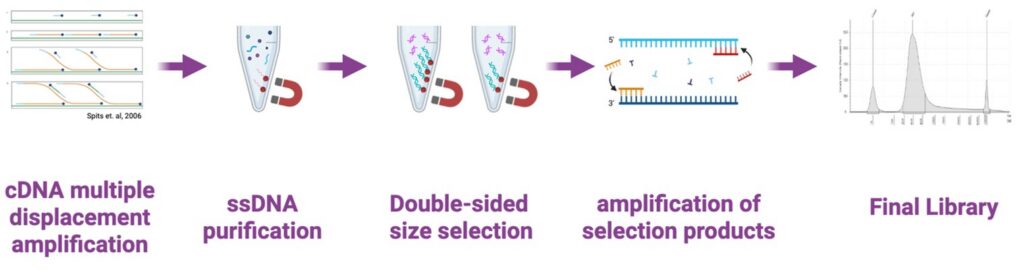
Following RNA capture, the bound transcripts are converted into complementary DNA (cDNA) and prepared into sequencing libraries using the Stereo-seq visualization reagent set (PE75). These libraries are then sequenced using high-throughput sequencing platforms. During sequencing, both the gene identity and the spatial barcode information are read, enabling the reconstruction of spatial transcriptomic profiles.
Data Analysis and Visualization
Finally, the sequencing data are processed using computational pipelines such as SAW and StereoMap. These tools map sequencing reads back to their spatial coordinates and generate high-resolution maps of gene expression across the tissue. Researchers can then visualize spatial patterns of gene activity, identify cell types and functional regions, and study interactions between cells and tissues.